38 diagram of human heart with labels
› print › heart-diagramsHeart Diagram – 15+ Free Printable Word, Excel, EPS, PSD ... Teachers and students use the heart diagram, in biological science, to study the structure and functions of a human being’s heart. Friends and colleagues on the other hand may find this diagram template useful when it comes to sending special, personalized gifts to their family members and significant others. Download the template today, and ... › 1-label-the-heartLabel the heart — Science Learning Hub Label the heart Add to collection In this interactive, you can label parts of the human heart. Drag and drop the text labels onto the boxes next to the diagram. Selecting or hovering over a box will highlight each area in the diagram. Pulmonary vein Right atrium Semilunar valve Left ventricle Vena cava Right ventricle Pulmonary artery Aorta
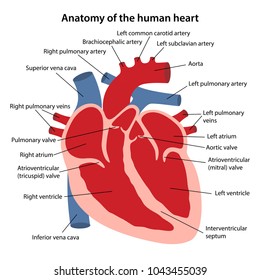
Heart Diagram with Labels and Detailed Explanation The heart is located under the ribcage, between the lungs and above the diaphragm. It weighs about 10.5 ounces and is cone shaped in structure. It consists of the following parts: Heart Detailed Diagram Heart - Chambers There are four chambers of the heart . The upper two chambers are the auricles and the lower two are called ventricles.

Diagram of human heart with labels
Diagram of Human Heart and Blood Circulation in It | New ... Exterior of the Human Heart A heart diagram labeled will provide plenty of information about the structure of your heart, including the wall of your heart. The wall of the heart has three different layers, such as the Myocardium, the Epicardium, and the Endocardium. Here's more about these three layers. Epicardium byjus.com › biology › human-heartHuman Heart - Anatomy, Functions and Facts about Heart The human heart is one of the most important organs responsible for sustaining life. It is a muscular organ with four chambers. The size of the heart is the size of about a clenched fist. The human heart functions throughout a person’s lifespan and is one of the most robust and hardest working muscles in the human body. commons.wikimedia.org › wiki › File:Diagram_of_theFile:Diagram of the human heart (cropped).svg - Wikimedia Apr 05, 2022 · English: Diagram of the human heart 1. Superior vena cava 2. 4. Mitral valve 5. Aortic valve 6. Left ventricle 7. Right ventricle 8. Left atrium 9. Right atrium 10. Aorta 11. Pulmonary v
Diagram of human heart with labels. PDF Anatomy of Heart Labeled and Unlabeled Images (a) Anterior view of the external heart C' 2019 Pearson Education. Aort'c arch Ligamentum arteriosum Left pulmonary artery Left pulmonary ve ns Auricle of left atrium Circumflex artery Left coronary artery (in atrioventricular sulcus) Great cardiac vein Left ventricle Anterior interventricular artery (in anterior interventricular sulcus) Apex Human Heart - Diagram and Anatomy of the Heart The heart is a muscular organ about the size of a closed fist that functions as the body's circulatory pump. It takes in deoxygenated blood through the veins and delivers it to the lungs for oxygenation before pumping it into the various arteries (which provide oxygen and nutrients to body tissues by transporting the blood throughout the body). Heart Labeled Stock Illustrations - 213 Heart Labeled ... Download 213 Heart Labeled Stock Illustrations, Vectors & Clipart for FREE or amazingly low rates! New users enjoy 60% OFF. 186,012,893 stock photos online. How to Draw the Internal Structure of the Heart (with ... To draw the internal structure of a human heart, follow the steps below. Part 1 Finding a Diagram 1 To find a good diagram, go to Google Images, and type in "The Internal Structure of the Human Heart". Find an image that displays the entire heart, and click on it to enlarge it. 2 Find a piece of paper and something to draw with.
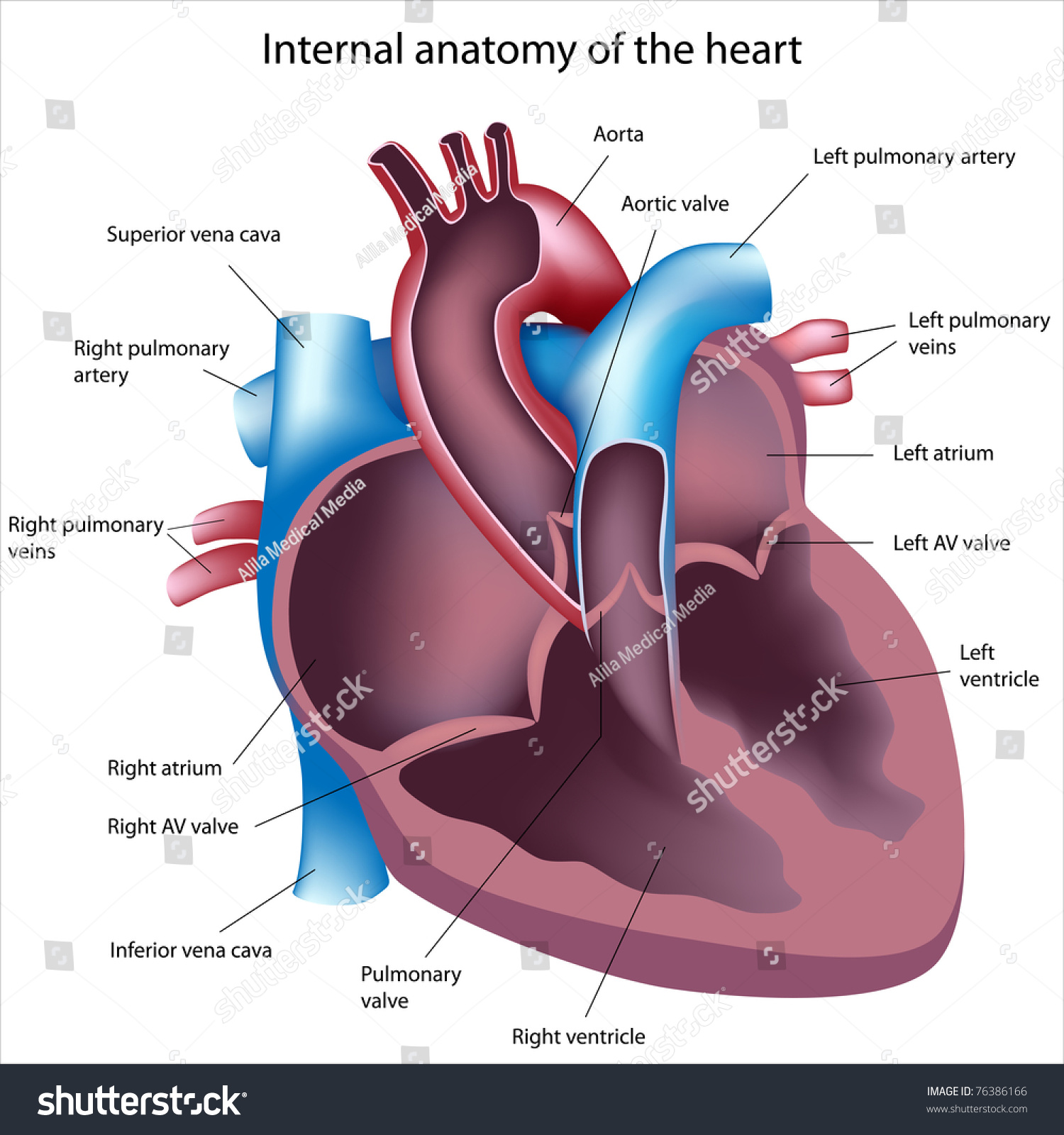
Free Printable Heart Diagram for Kids - Labeled and ... Oct 17, 2018 - Learn all the parts of the human heart by memorizing this free printable human heart diagram. Includes labeled and unlabeled versions. en.wikipedia.org › wiki › File:Diagram_of_the_humanFile:Diagram of the human heart (cropped).svg - Wikipedia File:Diagram of the human heart (cropped).svg. Size of this PNG preview of this SVG file: 611 × 600 pixels. Other resolutions: 244 × 240 pixels | 489 × 480 pixels | 782 × 768 pixels | 1,043 × 1,024 pixels | 2,086 × 2,048 pixels | 663 × 651 pixels. This is a file from the Wikimedia Commons. Information from its description page there is ... How to draw human heart labeled | Human drawing, Heart ... How to draw human heart labeled. External structure of human heart shows its conical shape with apex facing downwards and the broad base directed upwards. It is enclosed in double layered, transparent, thin sac called "Pericardium" . The space between the inner and outer layers is called Pericardial space, it is filled with pericardial fluid ... Label Heart Interior Anatomy Diagram - Enchanted Learning Label Heart Interior Anatomy Diagram: Human Anatomy: The heart is a fist-sized, muscular organ that pumps blood through the body. Oxygen-poor blood enters the right atrium of the heart (via veins called the inferior vena cava and the superior vena cava). The blood is then pumped into the right ventricle and then through the pulmonary artery to ...
Human Heart Diagram - Human Body Pictures - Science for Kids Photo description: This is an excellent human heart diagram which uses different colors to show different parts and also labels a number of important heart component such as the aorta, pulmonary artery, pulmonary vein, left atrium, right atrium, left ventricle, right ventricle, inferior vena cava and superior vena cava among others. Human Heart: Label the diagram 1 worksheet Human Heart: Label the diagram 1 Study the figure carefully.Label the 10 parts of the human heart A-J. 13+ Heart Diagram Templates - Sample, Example, Format ... Free heart diagrams can be helpful for students in understanding the heart and its functioning. Human heart is a complicated figure and for students from science, they will often need the images of the heart for its illustration. The above collection of heart samples will make it easier for students to download, print and use it in their projects. Understanding Human Heart with Heart Diagram | EdrawMax Online 3.2 How to Draw A Heart Diagram in EdrawMax. Step 1: Open EdrawMax, and select Science and Education, then click on human organs. Step 2: Use the wide range of symbols from the libraries available to create your heart diagram. Step 3: Add in your text and label the heart diagram to suit the requirements.
31 Blood Vessels Diagram To Label
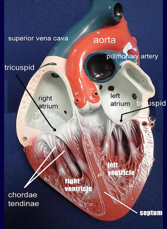
The Human Heart Labeling Worksheet (Teacher-Made) Two lower chambers - the left and right ventricles. It's also made up of four valves - these are known as the tricuspid, pulmonary, mitral and aortic valves. With this heart diagram without labels, you can familiarise your students with all the correct terms and help them recognise all these features of the anatomy.
Label Heart Diagram - Human Anatomy
byjus.com › biology › diagram-of-heartHeart Diagram with Labels and Detailed Explanation - BYJUS The human heart is the most crucial organ of the human body. It pumps blood from the heart to different parts of the body and back to the heart. The most common heart attack symptoms or warning signs are chest pain, breathlessness, nausea, sweating etc. The diagram of heart is beneficial for Class 10 and 12 and is frequently asked in the ...
Heart Models
Human Heart Diagram Labeled - Science Trends Let's examine the anatomy of the heart along with some diagrams that show how the heart operates. Anatomy Of The Heart The human heart usually weighs somewhere between 10 to 12 ounces in men and between 8 to 10 ounces in women, and in terms of size is roughly the size of the fist.

Label Kidney Diagram - Human Anatomy
› male-human-anatomy-diagramMale Human Anatomy Diagram Pictures, Images and Stock Photos Pacemaker Diagram Cross section of a human heart with pacemaker fitted, showing the major arteries and veins. This is an EPS 10 vector illustration and includes a high resolution JPEG. male human anatomy diagram stock illustrations
Anatomy Review: The Heart
File:Diagram of the human heart (no labels).svg ... File:Diagram of the human heart (no labels).svg. Size of this PNG preview of this SVG file: 498 × 599 pixels. Other resolutions: 199 × 240 pixels | 399 × 480 pixels | 499 × 600 pixels | 639 × 768 pixels | 851 × 1,024 pixels | 1,703 × 2,048 pixels | 533 × 641 pixels.
labeled heart Gallery
PDF Free Anatomy Coloring Page - NCSU Heart this drawing shows how Olcod 'lows through the heart. Color Me. The ate.2S the heart With oxygen ate labeled with at'l Color these areas The areas o' the heart with less oxygen ate labeled with a color areas BLUE. ARTERY LEFT LUNG PULMONARY VEINS AORTA PULMONARY VEINS raGHT LUNG ATRIUM RIGHT VENTRICLE INFERIOR VFNACAVA LEFT LEFT VENTRICLE

Dentistry and Medicine: Thorax,Lungs,Heart Anatomy and Physiology Diagrams Free Download
Human Heart Diagram Photos and Premium High Res Pictures ... heart illustration pulmonary artery kidney diagram 612 Human Heart Diagram Premium High Res Photos Browse 612 human heart diagram stock photos and images available, or search for heart illustration or pulmonary artery to find more great stock photos and pictures. of 11 NEXT
Respiratory System Worksheet - WikiEducator
A Labeled Diagram of the Human Heart You Really Need to ... The human heart, comprises four chambers: right atrium, left atrium, right ventricle and left ventricle. The two upper chambers are called the left and the right atria, and the two lower chambers are known as the left and the right ventricles. The two atria and ventricles are separated from each other by a muscle wall called 'septum'.
Post a Comment for "38 diagram of human heart with labels"