38 microscope images with labels
en.wikipedia.org › wiki › Electron_microscopeElectron microscope - Wikipedia An electron microscope is a microscope that uses a beam of accelerated electrons as a source of illumination. As the wavelength of an electron can be up to 100,000 times shorter than that of visible light photons , electron microscopes have a higher resolving power than light microscopes and can reveal the structure of smaller objects. abcnews.go.com › technologyTechnology and Science News - ABC News Jul 12, 2022 · The telescope was launched in December 2021 and sent back the first full-color images from its orbit 1 million miles from Earth. July 12 Pig organ transplants inch closer with testing in the dead
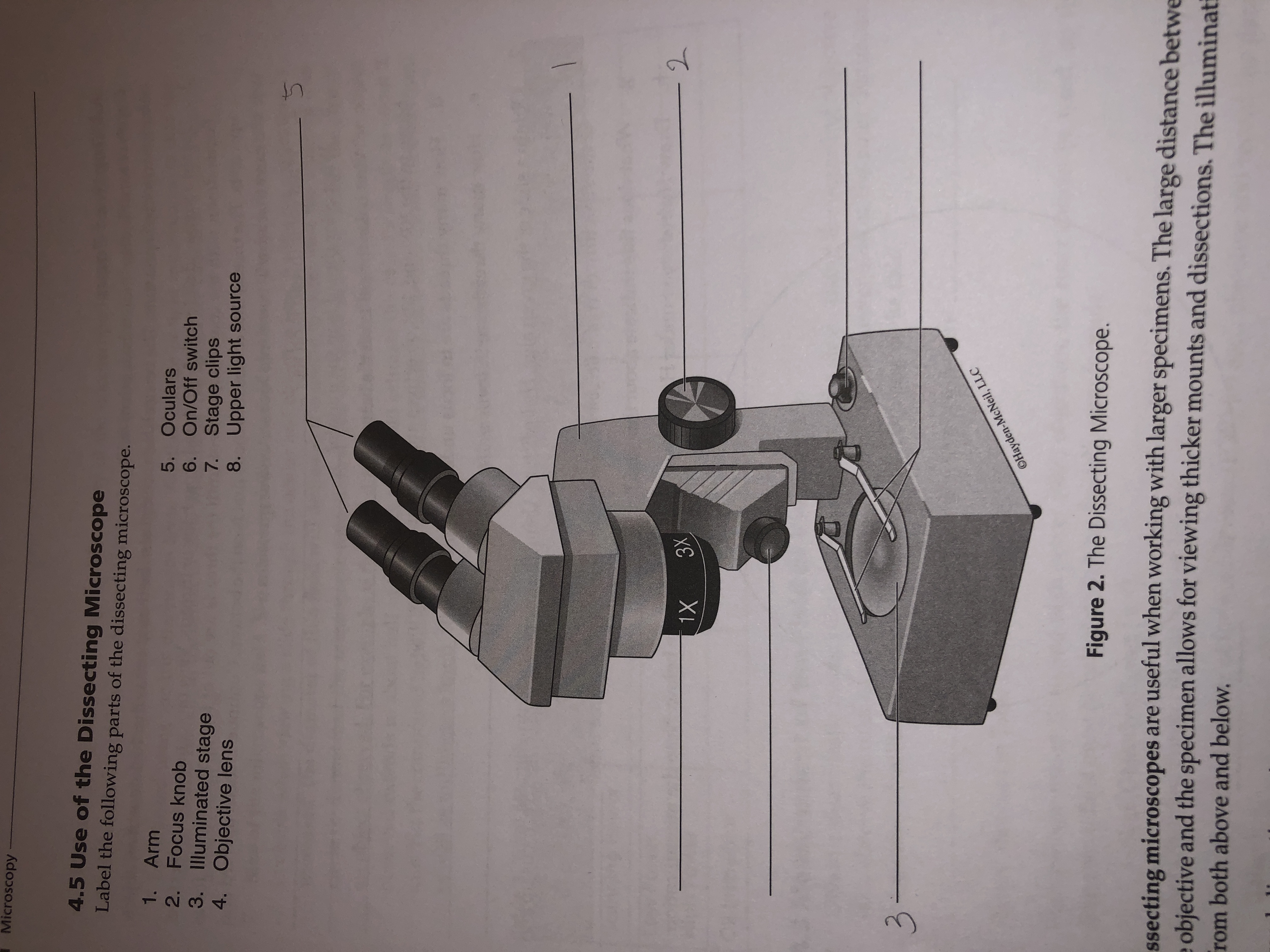
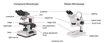
rsscience.com › stereo-microscopeParts of Stereo Microscope (Dissecting microscope) – labeled ... Unlike a compound microscope that offers a flat image, stereo microscopes give the viewer a 3-dimensional image that you can see the texture of a larger specimen. [In this image] Examples of Stereo & Dissecting microscopes. Major microscope brands (Zeiss, Olympus, Nikon, Amscope, Omano, Leica …) all produce stereomicroscopes.
Microscope images with labels
› microscopy › enZEISS LSM 980 with Airyscan 2 – Confocal Microscope with ... Specific areas of interest were imaged with the Airyscan 2 detector in order to acquire high resolution images of the Purkinje cells. The Airyscan 2 datasets were processed and orthogonal projections were created with ZEN Blue. The individual superresolution images were aligned with the cerebellum using ZEN Connect. en.wikipedia.org › wiki › Scanning_electron_microscopeScanning electron microscope - Wikipedia A scanning electron microscope (SEM) is a type of electron microscope that produces images of a sample by scanning the surface with a focused beam of electrons.The electrons interact with atoms in the sample, producing various signals that contain information about the surface topography and composition of the sample. › products › microscopeMicroscope Objective Lens | Products | Leica Microsystems The objective lens is a critical part of the microscope optics. The microscope objective is positioned near the sample, specimen, or object being observed. It has a very important role in imaging, as it forms the first magnified image of the sample. The numerical aperture (NA) of the objective indicates its ability to gather light and largely determines the microscope’s resolution, the ...
Microscope images with labels. › books › NBK26880Looking at the Structure of Cells in the Microscope ... In electron-microscope (EM) tomography, the specimen holder is tilted in the microscope, which achieves the same result. In this way, one can arrive at a three-dimensional reconstruction, in a chosen standard orientation, by combining a set of views of many identical molecules in the microscope's field of view. › products › microscopeMicroscope Objective Lens | Products | Leica Microsystems The objective lens is a critical part of the microscope optics. The microscope objective is positioned near the sample, specimen, or object being observed. It has a very important role in imaging, as it forms the first magnified image of the sample. The numerical aperture (NA) of the objective indicates its ability to gather light and largely determines the microscope’s resolution, the ... en.wikipedia.org › wiki › Scanning_electron_microscopeScanning electron microscope - Wikipedia A scanning electron microscope (SEM) is a type of electron microscope that produces images of a sample by scanning the surface with a focused beam of electrons.The electrons interact with atoms in the sample, producing various signals that contain information about the surface topography and composition of the sample. › microscopy › enZEISS LSM 980 with Airyscan 2 – Confocal Microscope with ... Specific areas of interest were imaged with the Airyscan 2 detector in order to acquire high resolution images of the Purkinje cells. The Airyscan 2 datasets were processed and orthogonal projections were created with ZEN Blue. The individual superresolution images were aligned with the cerebellum using ZEN Connect.
Answered: Microscopy 4.5 Use of the Dissecting… | bartleby

Label The Microscope Parts! Diagram | Quizlet
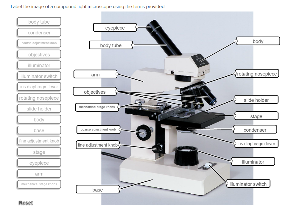
Solved Label the image of a compound light microscope using ...

This is a common compound microscope Label its parts class 11 ...
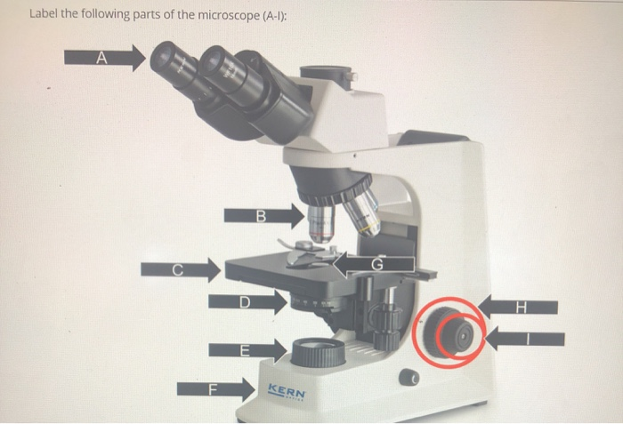
Solved Label the following parts of the microscope (A-1 ...
Parts of a Microscope with Their Functions – Microbe Online

Microscope Labels I - L Diagram | Quizlet

Microscope Terms Glossary | Earth science lessons, Biology ...

Meiji MT6500 Series PCM NIOSH 7400 Asbestos Microscope ...

Simple Microscope - Diagram (Parts labelled), Principle ...
Microscope Review Name ______ Date_______ Part 1. Label the ...
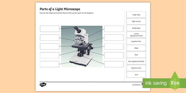
Parts of a Light Microscope Activity | Labeling Task
Microscope labeled diagram

Cytology - BIO 1210: Human Anatomy and Physiology I ...

Microscope Diagram Labeled, Unlabeled and Blank | Parts of a ...

Microscope Labeling Diagram | Quizlet
Transmitted light microscope B3 Professional series B3-220ASC ...

MICROSCOPE Labeling - Part - 3

Label a Microscope Worksheet by NC Middle School Resources | TpT
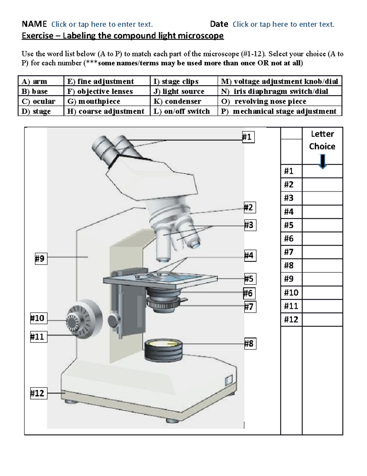
BIO 101 parts of the microscope to label - NAME Click or tap ...

label microscope diagram | Charts | Microscope, Anatomy bones ...
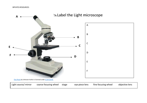
Label the light microscope | Teaching Resources
Getting Started - Virtual Fluorescent Microscope - Wartburg ...

Microscope Label Diagram | Quizlet

Dissecting Stereo Microscope Parts and Functions
Parts of Stereo Microscope (Dissecting microscope) – labeled ...

A Study of the Microscope and its Functions With a Labeled ...

Microscope Labeling Activity - SMART Board Activity ...

Labeling a Microscope Free Worksheet Pack

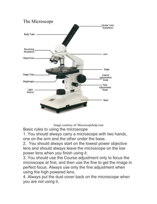
Parts of the Microscope with Labeling (also Free Printouts ...
Parts of a Microscope with Their Functions – Microbe Online
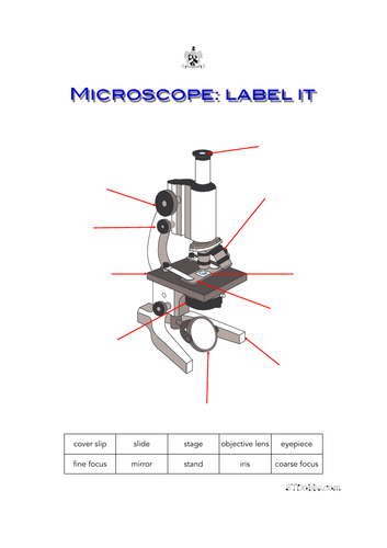
Microscope: label it | Teaching Resources
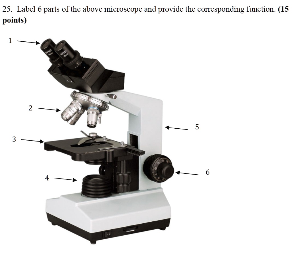
SOLVED: 25. Label 6 parts of the above microscope and provide ...

Label the numbered parts of the microscope - ppt download

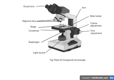
Compound Microscope: Definition, Diagram, Parts, Uses ...

Parts of a Microscope Labeling Activity

Microscope With Labels clip art | Microscope parts ...
Microscope With Labels Clip Art-vector Clip Art-free Vector ...
Post a Comment for "38 microscope images with labels"